
Anesthesia12 min read
Complete heart block in a cat who was booked for surgery.
Smokey came in for a mass removal, not a cardiology work up. He was bright, active, and had no clinical signs at home. His atria and ventricles were beating completely independently of each other, and that changes how you anesthetize him.

TopicsComplete heart blockThird-degree AV blockFelineBradyarrhythmiaAV dissociationAnesthesia riskPacing
Clinical presentation.
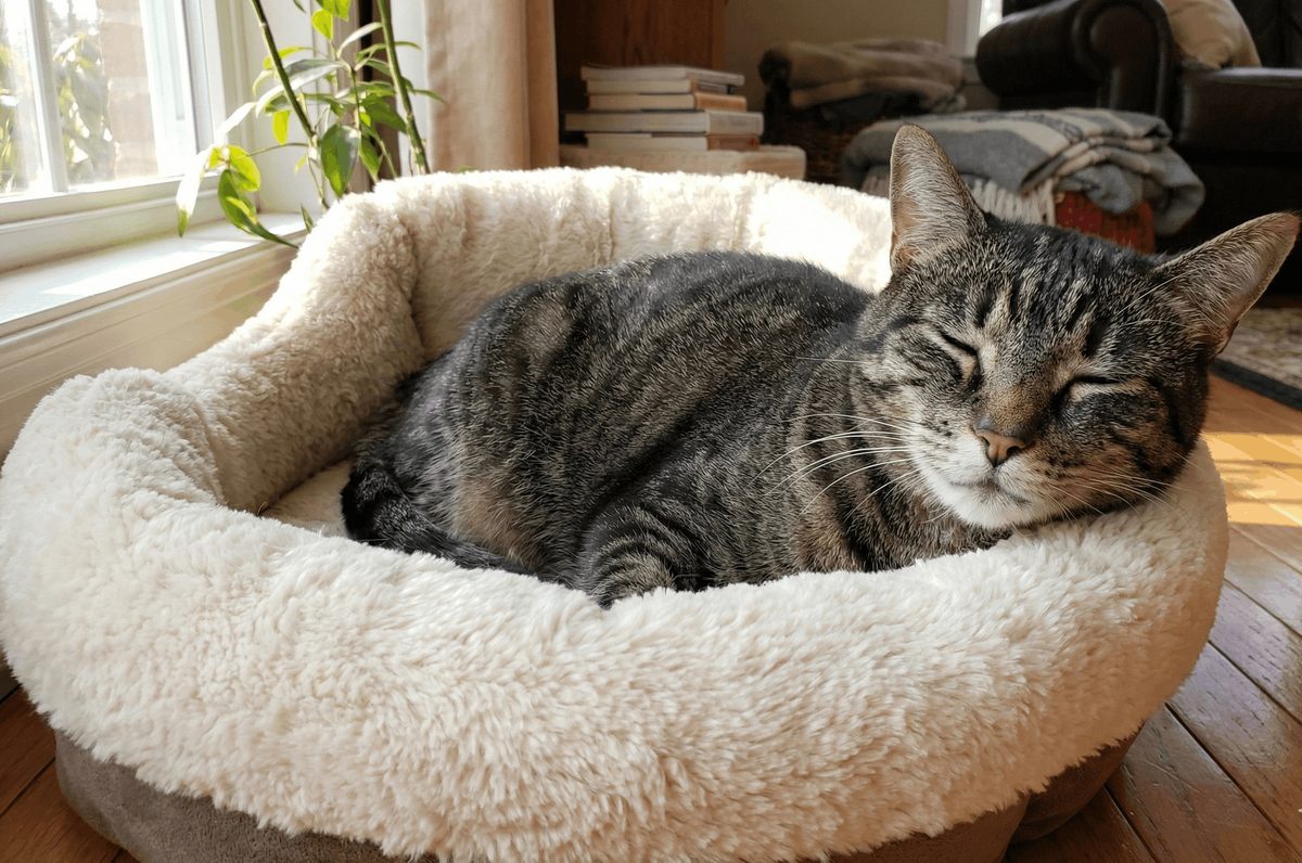
“Smokey,” 12-year-old male neutered Domestic Short Hair
- History: Presented for scheduled surgical removal of a sublingual mass, with a prior diagnosis of atrioventricular block, complete heart block in his case.
- At home: Doing very well. No clinical signs reported, reasonably active, no concerns noted by the owner.
Smokey did not come to me because of his heart. He came in because he had a sublingual mass that needed to come out, and somewhere along the way an atrioventricular block had been picked up. At home, as far as his owner was concerned, he was a normal twelve-year-old cat.
That combination, a cat who looks completely well and a heart that is in complete block, is exactly why this case is worth walking through. Because the moment you plan to anesthetize him, the rhythm stops being an incidental finding.
Why an asymptomatic cat is still a problem.
Complete heart block, or third degree AV block, is uncommon in cats, but when you find it, it usually indicates intrinsic conduction system disease.
Cats with complete heart block may appear asymptomatic at home. This is because they tend to have faster escape rhythms, particularly when compared with dogs. In dogs we will usually see clinical signs suggestive of reduced cardiac output, things like lethargy, weakness, reduced activity or exercise tolerance, and in some patients syncope or collapse, especially with the lower escape rates.
The anesthetic problem
- General anesthesia poses increased risk in these patients, because we can see suppression of the escape pacemakers.
- This can result in an increased risk of profound bradycardia or asystole.
- These patients are often considered at higher anesthetic risk.
So a cat who is thriving on the sofa can still be a cat who arrests on induction. That is the whole tension in Smokey's case.
Normal conduction, briefly.
It is worth reviewing normal sinus rhythm first, because complete heart block only makes sense once you can see what it interrupts.
Up in the high right atrium we have the sinoatrial node, which is the dominant pacemaker of the heart. It depolarizes and delivers a wave of depolarization spreading through the atria, and that results in the P wave on the ECG. The impulse then enters the AV node, which slows it down, and delivers it through to the His-Purkinje system, where the ventricles depolarize.
Now remember that the heart has a fibrous skeleton, and this electrically insulates the atria from the ventricles. The AV junction is the only normal electrical bridge between the two.
What complete heart block actually is.
If that bridge fails completely, the atria and the ventricles must function independently. There is complete electrical disconnection between them, and no atrial impulses, zero, reach the ventricles. That is complete heart block, and ventricular activation becomes independent of the atrial rhythm.
The sinus node carries on doing its job perfectly well. It keeps depolarizing the atria, so you keep seeing P waves. Every one of those impulses is then blocked at the AV junction.
So why does the heart not just stop?
Because the ventricles survive by relying on what we call subsidiary pacemakers. Two possibilities exist, and which one you get depends on how distal the block is.
| Junctional escape rhythm | Ventricular escape rhythm |
|---|---|
| The His bundle or proximal junctional tissue remains capable of automaticity. | The junctional tissue cannot generate impulses, so the ventricular myocardium becomes the pacemaker. |
| Ventricular activation still uses the normal His-Purkinje system. | Ventricular activation is slow and disorganized, spreading by cell to cell conduction rather than through the normal His-Purkinje system. |
| QRS complexes are narrow. | QRS complexes are wide and bizarre. |
| Typically what we see in cats. | Suggests a focus lower down in the ventricles. |
Subsidiary pacemakers in complete AV block
Smokey's ECG.
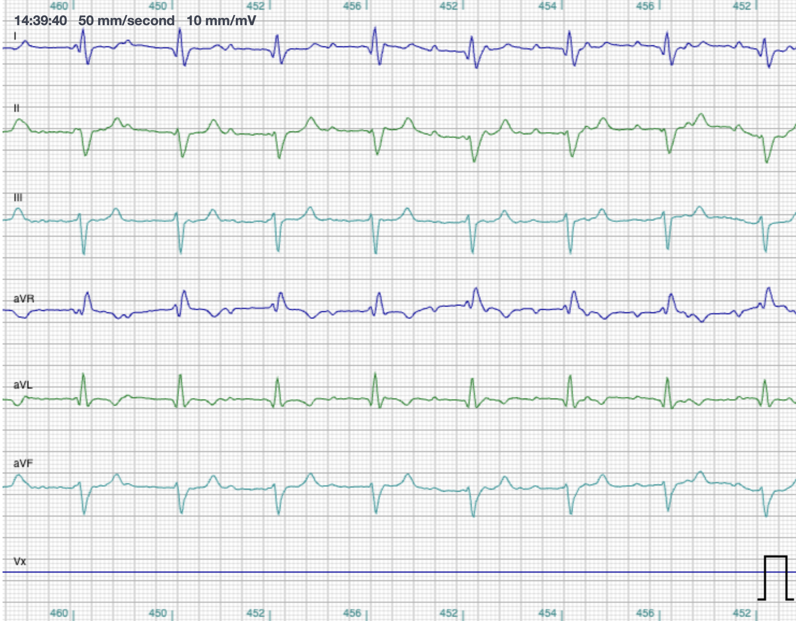
- Ventricular rate: the R to R interval is about 138 beats per minute, and it is regular.
- Atrial rate: the P to P interval is about 240 beats per minute, and it is regular.
- Relationship: there are more P waves than QRS complexes, and there is no consistent relationship between them. The P waves simply march through the ECG without any fixed PR interval.
- QRS: on the wider side, with a deviation in the axis.
- Interpretation: I suspect this cat has a junctional escape rhythm, which can have a slightly prolonged QRS duration, and the lead findings look to be consistent with left anterior fascicular block, which is a common change we see in cats, often associated with HCM.
Work through it in that order and the diagnosis assembles itself. Two rhythms, each perfectly regular on its own. The atria going at roughly 240, the ventricles going at roughly 138. More P waves than QRS complexes. And critically, no fixed relationship between the two. The atrial rhythm and the ventricular rhythm are independent of each other.
That is atrioventricular dissociation, and here it is caused by complete block.
Complete heart block (third degree AV block) with a junctional escape rhythm.
The same block, a very different escape.
Compare Smokey with this dog, who has a slower ventricular escape focus. Here you see the wide QRS complexes, and the P waves marching through completely dissociated, indicating third degree heart block with a ventricular escape focus.
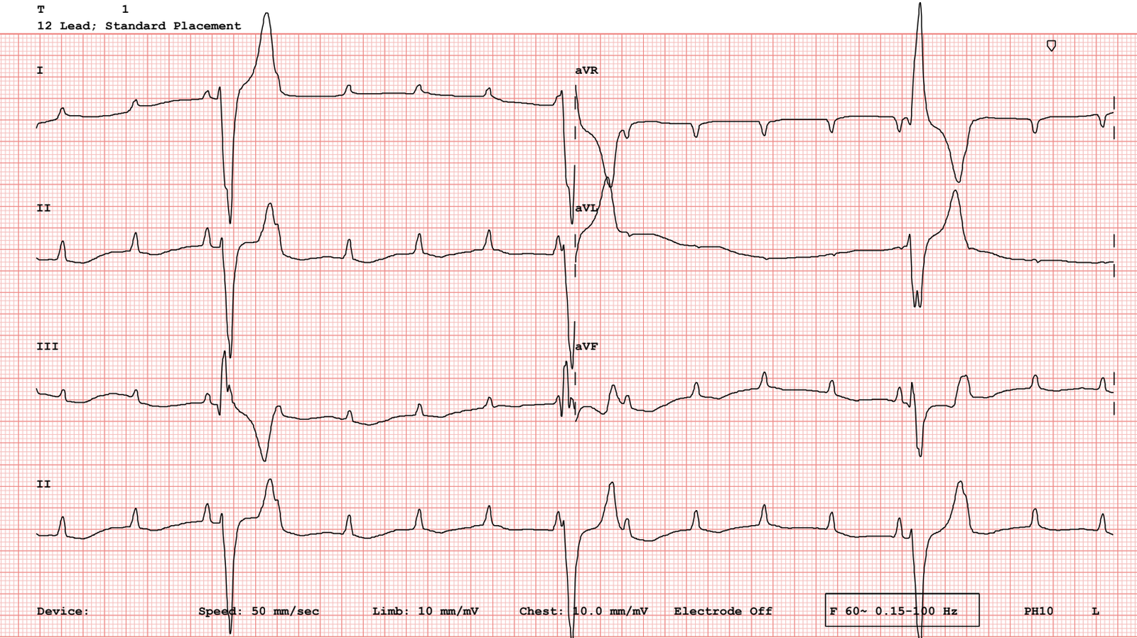
There is one finding here that surprises people, and it is worth knowing. Unlike in humans, in studies in dogs with high grade or complete AV block, narrower escape rhythms have been shown to be associated with a poor prognosis, while wider, slow ventricular escape rhythms are associated with longer survival. That is not what is described in the human literature.
Mechanism.
- There is typically severe damage to the AV conduction system in patients with complete heart block.
- In cats, most commonly, we suspect some sort of degeneration or fibrous replacement of that conduction system, in the AV node and bundle of His area.
- It has also been reported in patients with hyperthyroidism, and in those with underlying cardiomyopathies.
Differential diagnoses.
Before you settle on complete heart block, these are the causes of AV dissociation and severe bradycardia in dogs and cats that need ruling out.
| Atrial standstill | Severe hyperkalemia | Atrial fibrillation with complete heart block |
|---|---|---|
| No P waves at all, but there will be a junctional or ventricular escape focus. | Blunting of the P wave, leading to no atrial activity, and the QRS becomes widened. A life-threatening but reversible cause of bradyarrhythmia. | A slow, regular ventricular escape rhythm, but with chaotic atrial activity or fibrillatory waves present. |
Differential diagnoses for AV dissociation and severe bradycardia
That last one comes with a caveat in this species. Fibrillatory waves can be hard to see on cat ECGs because of the low amplitude, and it can end up looking very similar to atrial standstill.
Diagnostics.
- General laboratory work, so biochemistry and hematology, and of course electrolytes.
- Thyroid testing in cats.
- Echocardiography, to evaluate for evidence of structural heart disease, and for things like bradycardia-induced cardiomyopathy, where we might see dilation of the heart chambers.
- Chest radiographs for screening, or if there is a clinical suspicion of congestive heart failure or impending congestive heart failure.
Smokey's echo.
You can see his arrhythmia on the ECG trace running beneath the echo image. His heart structure and function, though, looked reasonably normal.
Echocardiogram
- No overt HCM phenotype.
- Left atrial size looked pretty normal.
- Left ventricular systolic function looked pretty normal and appropriate.
Anesthesia planning.
So what do we do with a cat who is seemingly asymptomatic at home, but needs to have a surgical procedure done? The priority is stabilization for surgery.
- Cats with complete heart block should be considered a high anesthetic risk.
- We usually recommend that these patients have surgery and anesthesia performed at a facility where there is access to temporary pacing systems, and the ability to go ahead with permanent pacing if necessary.
- These things should be on hand prior to induction, as the induction agents may precipitate severe bradycardia and cardiovascular collapse.
When does a cat with complete heart block need a pacemaker?
Usually when the patient is symptomatic, or when the bradyarrhythmia causes significant hemodynamic compromise.
- Cats exhibiting clinical signs such as syncope, weakness, or collapse. Syncope has been reported in anywhere between about 20 and the high 50 percents of cats with high grade AV block.
- Cats tend to maintain an escape rate in the range of 100, 120, to 140 beats per minute, but this can diminish over time. We are usually thinking about a pacemaker when the patient becomes symptomatic, and when the ventricular escape rate is dropping below 100 beats per minute.
- These chronic bradyarrhythmias can also result in bradycardia-induced volume overload and the subsequent development of congestive heart failure. Preventing and managing that progression is an indication for pacing.
This is where cats and dogs differ. Unlike in dogs, where third degree AV block is frequently fatal without pacing, cats can often tolerate the arrhythmia well for extended periods without intervention. A high proportion of cats with third degree AV block have a junctional escape rhythm with narrow QRS complexes and a relatively rapid rate, over 100 and usually up to 140 beats per minute. Because that rate often supports adequate cardiac output, clinical signs are less common, and many cats can survive beyond a year without specific therapy.
Because of that better natural prognosis, pacemaker implantation is rarely performed in cats when compared with dogs. When it is necessary, in those symptomatic cats, epicardial pacing, meaning surgical placement of the leads on the myocardium, is what we use in our feline patients in most cases.
What happened to Smokey.
His echo showed largely normal looking structure and function, with no overt HCM phenotype. So he went ahead and had his surgery.
- We placed transthoracic external pacing pads prophylactically prior to induction.
- He remained stable throughout the procedure, and there was no requirement for pacing.
- His escape rhythm remained stable.
- His mass was excised without any complications.
- He recovered well from surgery and went home the following day.
As far as we are aware, he continues to do well at home.
Summary.
- Complete heart block is identified by independent atrial and ventricular rhythms, otherwise termed AV dissociation.
- In cats, unlike in dogs, the ventricular escape rate is often fast enough that clinical signs might be masked or absent.
- The block is frequently attributed to irreversible change or degeneration of the AV conduction system, although we do need to screen for other disease processes.
- Even in patients that are asymptomatic and outwardly stable, complete heart block carries an elevated risk for anesthesia, and temporary or permanent pacing capabilities must be available at the facility for surgery.
- Definitive treatment for symptomatic patients is permanent pacemaker implantation.
Common questions about complete heart block.
What is complete heart block in a cat?
Complete heart block, or third degree AV block, is a total failure of conduction at the AV junction. The atria and the ventricles become electrically disconnected, and no atrial impulses at all reach the ventricles. The sinus node carries on depolarizing the atria, so P waves keep appearing, but each one is blocked. The ventricles are kept going by a subsidiary pacemaker, and ventricular activation becomes completely independent of the atrial rhythm.
How do you recognize it on the ECG?
Look for two independent rhythms. The P waves are regular. The QRS complexes are regular. There are more P waves than QRS complexes, and there is no consistent relationship between them, so the P waves march through the trace with no fixed PR interval. In Smokey's ECG, the atrial rate was about 240 beats per minute and the ventricular rate about 138, each entirely regular on its own.
Why do cats with complete heart block often look normal at home?
Because they tend to have faster escape rhythms than dogs do, often in the range of 100 to 140 beats per minute. That rate is frequently enough to support adequate cardiac output, so clinical signs may be subtle or absent altogether. Dogs, whose escape rates are typically slower, are far more likely to show lethargy, weakness, reduced exercise tolerance, and syncope or collapse.
How do you tell a junctional escape from a ventricular escape?
Look at the width of the QRS. If the His bundle or proximal junctional tissue is still capable of automaticity, ventricular activation continues to use the normal His-Purkinje system, so the QRS complexes are narrow. That is a junctional escape, and it is typically what we see in cats. If the junctional tissue cannot generate impulses, the ventricular myocardium takes over, activation spreads slowly by cell to cell conduction, and the QRS complexes become wide and bizarre. That is a ventricular escape, and it points to a focus lower down.
Does a cat with third degree AV block need a pacemaker?
Usually only when the cat is symptomatic, or when the bradyarrhythmia is causing significant hemodynamic compromise. Syncope, weakness, and collapse are the signs that prompt it, and the escape rate dropping below 100 beats per minute is a common trigger for the conversation. Chronic bradyarrhythmia can also cause bradycardia-induced volume overload and congestive heart failure, and preventing that is itself an indication. Unlike dogs, where third degree AV block is frequently fatal without pacing, many cats tolerate it for extended periods, and pacemaker implantation is rarely performed in cats by comparison.
Why is anesthesia so risky in these patients?
Because the ventricles are depending entirely on an escape pacemaker, and anesthetic agents can suppress it. That brings a real risk of profound bradycardia or asystole on induction. A cat who is completely stable at home is still a high anesthetic risk, which is why these procedures should happen where temporary pacing systems are available and permanent pacing is possible, with everything on hand before induction rather than after something goes wrong.
What else can look like complete heart block?
Atrial standstill, where there are no P waves at all but there is still a junctional or ventricular escape focus. Severe hyperkalemia, where the P wave is blunted until atrial activity disappears and the QRS widens, which is life-threatening but reversible. And atrial fibrillation with complete heart block, which gives a slow regular escape rhythm alongside chaotic atrial activity. That last one is tricky in cats, because fibrillatory waves are low amplitude and can end up looking very like atrial standstill.
Get the full case breakdowns
Every case, in full, with the video walkthrough.
Each case breakdown on this site has a longer version. Dr. Hayley works through the ECG and the echo on video, the way she would talk a colleague through it. Subscribers get the full walkthrough with every case.
- The full video walkthrough with each case.
- New case breakdowns as they publish.
- Practical cardiology for vets and techs.
We respect your privacy.Privacy policy·Unsubscribe anytime.