
ECG12 min read
Premature atrial complexes in a Cavalier King Charles Spaniel with MMVD stage B1.
A routine wellness exam, a new murmur, and the occasional early beat. Molly's case is a good one to walk through, because the beat that interrupts her rhythm is one most vets and techs will meet again.

TopicsMyxomatous mitral valve diseaseCanineArrhythmiaMMVD stage B1Supraventricular arrhythmiaPremature atrial complex (PAC/APC)
Clinical presentation.

“Molly,” 5-year-old FS Cavalier King Charles Spaniel
- History: Presented for routine wellness exam. No concerns noted at home, recently noted heart murmur.
- Physical exam: Left apical systolic murmur (grade 3/6), regular rhythm with intermittent early beats. Normal pulmonary auscultation.
This is little Molly. She is a very cute five-year-old Cavalier King Charles Spaniel, and she came in for a routine wellness exam with nothing wrong as far as her owner was concerned. On physical exam I heard a left apical systolic murmur, grade three out of six. Her rhythm was predominantly regular, but there were occasional early beats audible. Everything else about her thoracic auscultation was normal.
There were two different findings here. First, the murmur. In a Cavalier that is close to expected, because we know MMVD is very common in this breed. Second, the occasional early beats, and those are the part that does not explain itself. A murmur tells you something about a valve. It tells you nothing about why the rhythm is being interrupted.
So we worked up both. The echo to look at the valve, and, because there was a rhythm abnormality on auscultation, an ECG to look at the beat.
Echo findings.
So we are going to take a look at her echo here. You can see there is some thickening of her mitral valve leaflets, and a little bit of prolapse. Her left atrium and left ventricle, though, look pretty normal in size in this view, and that is a detail to keep in mind as we go through this case.
Now let us take a look with color Doppler. There is mild mitral valve insufficiency, with a posteriorly directed jet of mitral regurgitation.
Echocardiogram
- Mild thickening and prolapse of the mitral leaflets, mild mitral and tricuspid regurgitation.
- Normal left atrial and left ventricular size, normal LV function.
Echo findings consistent with MMVD ACVIM stage B1
That puts her at ACVIM stage B1. There is disease, and there is no remodeling yet. Normal atrial size, normal ventricular size, normal function. That classification drives every management decision at the end of this case, so it is worth being sure of it before we go anywhere near the rhythm.
ECG findings.
Because I could hear the rhythm abnormality, we went ahead and did an ECG on her. When you look at Molly's strip, the underlying rhythm is predominantly sinus. It is just interrupted, every so often, by these premature complexes.
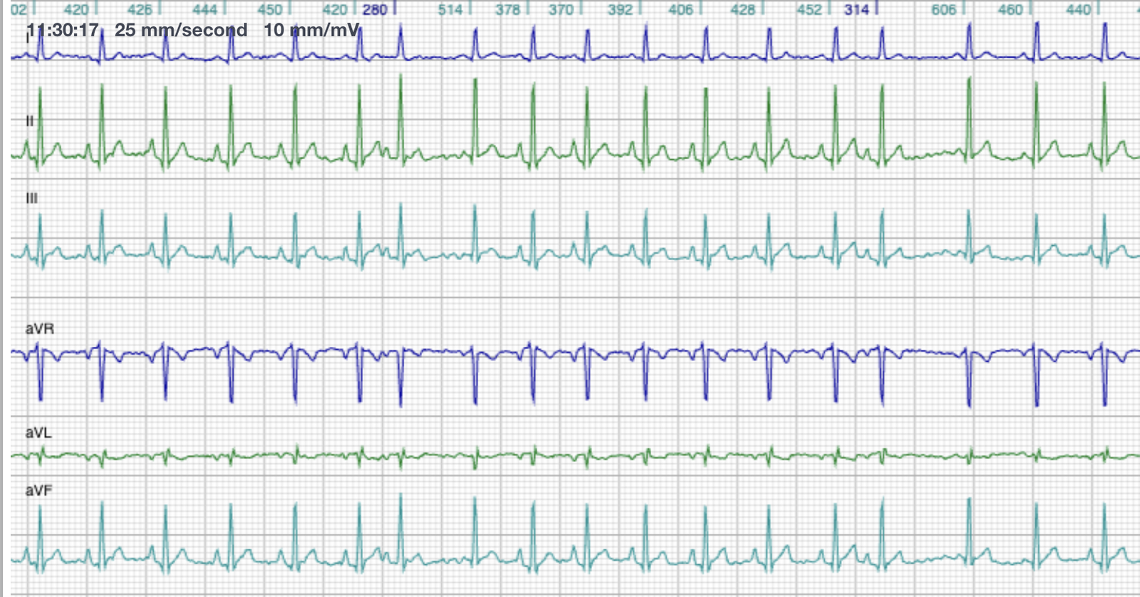
- Underlying rhythm: Sinus rhythm, HR ≈ 140 bpm.
- P waves:
– Sinus beats = normal morphology (positive in lead II)
– Occasional premature ectopic P waves (P′):
––> Lower amplitude but positive in II → ectopic atrial origin but close to sinus node
––> P–P′ interval shortened compared with baseline sinus P–P. - QRS: Narrow, normal morphology width (40ms (ref < 0.70 ms)); QRS follows premature P′.
- Pause: Non-compensatory (sinus node reset).
Those four lines are the whole diagnosis, and I want to walk you through them in the order I read them, because each one closes off a possibility.
First, the beats come earlier than the next expected sinus beat. That is what premature means here, and you can measure it: the P to P′ interval is shorter than the P to P interval of the normal sinus beats.
Second, if you take a look at the beats here, each premature one is preceded by a P′ wave that slightly differs in morphology from the sinus P waves. In Molly's case the P′ has a smaller amplitude. That difference is what tells you the impulse started somewhere else in the atria, at an ectopic focus, rather than at the sinoatrial node, which is the normal dominant pacemaker.

Third, take a look at the QRS complex following that ectopic P′. It looks normal and narrow in morphology, and it looks identical to the preceding sinus QRS complex. That tells you ventricular depolarization is still happening through the normal conduction system, so the beat is supraventricular in origin. It is coming from above the ventricles, not from within them, and that is what separates it from a ventricular premature complex.
And fourth, there is a pause before the next P wave is seen. This is what we call a non-compensatory pause, and it is typically what you see with premature atrial complexes. It is telling you the sinus node has been reset. Keep that pause in mind, because we are going to look at it more closely in a moment.
Sinus rhythm with occasional premature atrial complexes (PACs/APCs).
The electrophysiology of atrial premature beats.
Let me explore this a little further, because once you understand why sinus P waves and ectopic P waves look the way they do, you stop memorizing the pattern and start reading it. And reading it is what lets you work out a strip you have never seen before.
Remember that the sinoatrial node is the dominant pacemaker of the heart, and it sends out its wave of depolarization from there. The net electrical vector of that atrial depolarization points downward and leftward, toward the positive pole of lead II. And if you have a wave of depolarization moving toward the positive pole of lead II, you get a positive P wave in lead II. That is all a normal P wave is telling you.
So what happens if the beat starts from an ectopic atrial focus instead? Well, that focus could be in any number of different regions in the atria, and wherever it sits, the direction of the depolarization wave changes with it.
- Origin: Latent pacemakers or automatic foci from an atrial focus outside the sinus node.
- Timing: Occurs earlier than expected sinus impulse; may or may not reset SA node → non-compensatory pause or compensatory pause respectively.
- Conduction: Through AV node and His-Purkinje system → normal QRS.
- Depending on the origin of the ectopic focus, the direction of the atrial depolarisation wave will vary. Therefore: each origin yields a distinct P′ wave morphology..
- The P′ wave axis and shape provide diagnostic clues.
Which is exactly why Molly's P′ waves are worth a second look. They were lower in amplitude, but still positive in lead II, and that combination places her ectopic focus in the atria but close to the sinus node. The P′ morphology tells you roughly where the beat started.
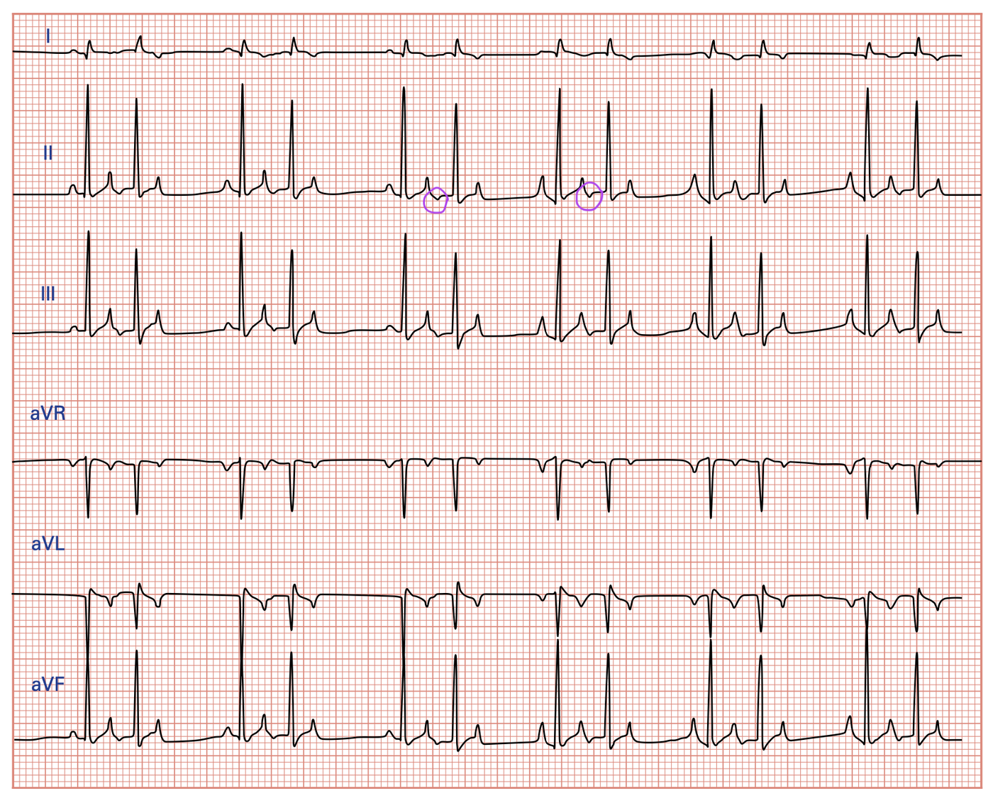
Compensatory vs non-compensatory pause.
That pause I asked you to keep in mind, this is why it matters. It is not dead space on the strip, it is evidence, and it answers one question. Did the ectopic beat reset the sinus node, or did the sinus node simply carry on regardless?
| Non-Compensatory Pause | Compensatory Pause | Interpolated APCs |
|---|---|---|
| Sinus node is reset by the ectopic depolarization | Sinus node continues on schedule (not reset) | No pause at all |
| (P–P′)+(P′–P) < 2x normal (P–P) interval | (P–P′)+(P′–P) = exactly 2 x normal (P–P) interval *Note – this can be complicated by sinus arrhythmia |
The ectopic P′ is inserted between two sinus beats |
| Most common pause type after PAC | Less common with atrial beats; more typical with VPCs | Often seen when APCs fall late in the cycle |
Pause Patterns Following APCs
Differential diagnoses.
Before you commit to PAC, run through the things that can look like one. This is the list I go through in my head, and it matters most on the strips that are not as tidy as Molly's.
| Sinus arrhythmia | Artifact | Atrial escape beat | APC with block | Premature junctional beat |
|---|---|---|---|---|
| Gradual rate variation with respiration → regular P morphology | Electrical noise or lead movement → no consistent P–QRS relationship | Occurs after pause, not premature | Ectopic P wave not followed by QRS (missed diagnosis if P buried in T wave) | Usually no P′ in front of narrow QRS (sometimes retrograde P′ with short P′R interval), may have hidden P′ or retrograde P′ evident in ST segment |
PAC: Differential diagnoses
What does this mean for Molly?
This is the question the owner in front of you is really asking, and for a dog like Molly the answer is reassuring.
- PACs are common and often benign.
- May occur in healthy animals or be associated with atrial stretch.
- Systemic/extracardiac stressors → catecholamines/metabolic stress (hypoxia, sepsis, uremia), electrolyte disturbance.
- Atrial inflammation, infiltration or compression.
Clinical significance in MMVD stage B1
- Early microscopic atrial changes
Mechanical stretch at the mitral annulus
Autonomic influences
Or incidental finding. - Usually isolated and clinically silent.
- Persistent or frequent PACs may predict later atrial fibrillation risk.
Management and follow up.
Molly does not need antiarrhythmic therapy, because her PACs are isolated. What she needs is a look for any triggers, and monitoring over time.
- No antiarrhythmic therapy needed if isolated.
- Address any triggers: pain, stress, electrolyte imbalance.
- Holter monitoring if clinical signs (exercise intolerance, syncope) or frequent PACs on resting ECG, especially if complexity present.
- Educate owners: watch for exercise intolerance, collapse, signs of CHF.
- MMVD Stage B1:
– No cardiac medications indicated
– No evidence that early pharmacologic therapy delays progression.
Prognosis.
And for a dog like Molly, the outlook is good.
- Stage B1 dogs with rare PACs → excellent prognosis, many dogs remain Stage B1 for years.
- Frequency of PACs often increases with progressive left-atrial enlargement (Stage B2 → C).
- Monitor: Re-examine (ECG and echo) q 6–12 mo.
Summary.
- MMVD, ACVIM stage B1 = murmur without chamber enlargement.
- Occasional PACs are common and often benign.
- PACs are diagnosed by: early timing, altered P′ morphology, narrow QRS, typically non-compensatory pause.
- No treatment necessary.

Questions that come up about PACs.
What is a premature atrial complex (PAC) in a dog?
A beat that arises from an ectopic focus in the atria rather than from the sinoatrial node, and that occurs earlier than the next expected sinus beat. On the ECG it is preceded by a P′ wave whose morphology differs from the sinus P waves, and the QRS that follows is narrow and identical to the sinus QRS, because conduction still travels through the AV node and His-Purkinje system. Also called an APC or an atrial premature beat, and classified as a supraventricular arrhythmia.
How do you tell a PAC from a VPC on the strip?
Look at the QRS, and at what comes before it. A PAC is preceded by an ectopic P′ wave and gives a narrow QRS identical to the sinus beats, because ventricular depolarization still happens through the normal conduction system. A ventricular premature complex arises below the AV node, so there is no preceding P′ and the QRS is wide and bizarre. The pause helps too: non-compensatory is the most common pause type after a PAC, while a compensatory pause is more typical with VPCs.
What is a non-compensatory pause, in one sentence?
It means the sinus node was reset by the ectopic beat, so the interval spanning the premature beat, (P–P′)+(P′–P), is less than twice the normal P–P interval. In a compensatory pause the sinus node carries on regardless and is not reset, so that same interval equals exactly twice the normal P–P interval. Sinus arrhythmia can complicate the measurement.
Why does the ectopic P wave look different?
Because the focus sits somewhere other than the sinus node, so the direction of the atrial depolarization wave changes. In sinus rhythm the net vector points downward and leftward, toward the positive pole of lead II, giving a positive P wave in lead II. Each ectopic origin yields a distinct P′ morphology, which is why the P′ axis and shape are diagnostic clues. Molly's P′ was lower in amplitude but still positive in lead II, placing the focus in the atria but close to the sinus node.
Are PACs dangerous in a dog?
They are common and often benign. They occur in healthy animals, and they are associated with atrial stretch, with systemic and extracardiac stressors such as hypoxia, sepsis, uremia and electrolyte disturbance, and with atrial inflammation, infiltration or compression. In stage B1 they are usually isolated and clinically silent. The reason to keep watching is that persistent or frequent PACs may predict later atrial fibrillation risk.
Does a dog with MMVD stage B1 and occasional PACs need medication?
No antiarrhythmic therapy is needed if the PACs are isolated, and triggers such as pain, stress, or electrolyte imbalance should be addressed instead. For the MMVD itself at stage B1 there are no cardiac medications indicated, and there is no evidence that early pharmacologic therapy delays progression. Holter monitoring earns its place if there are clinical signs such as exercise intolerance or syncope, or if there are frequent PACs on a resting ECG, especially with complexity present.
What is the prognosis?
Stage B1 dogs with rare PACs have an excellent prognosis, and many remain stage B1 for years. PAC frequency often increases as the left atrium enlarges and disease progresses toward stage B2 and C, which is why the recheck matters: ECG and echo every six to twelve months, and owners taught to watch for exercise intolerance, collapse, and signs of CHF.
Get the full case breakdowns
Every case, in full, with the video walkthrough.
Each case breakdown on this site has a longer version. Dr. Hayley works through the ECG and the echo on video, the way she would talk a colleague through it. Subscribers get the full walkthrough with every case.
- The full video walkthrough with each case.
- New case breakdowns as they publish.
- Practical cardiology for vets and techs.
We respect your privacy.Privacy policy·Unsubscribe anytime.