
ECG10 min read
An arrhythmia in a young dog before surgery.
Beansie came in to be spayed. On auscultation her rhythm was regularly irregular, and that one finding is enough to stop an anesthetic. The question is whether it needs treating, or whether it is a completely normal dog doing a completely normal thing.

TopicsRespiratory sinus arrhythmiaWandering pacemakerCanineNormal variantPre-anesthetic screeningAutonomic tone
Clinical presentation.
“Beansie,” 2-year-old female mixed breed dog
- History: Presented for a routine wellness exam and pre-anesthetic screening prior to being spayed.
- At home: Doing very well. No clinical signs reported by the owner. Normal exercise tolerance, no history of syncope, no coughing, normal breathing character.
- Physical examination: Bright, alert, and responsive. Pink mucous membranes, capillary refill time less than two seconds. Optimal body condition score. Pulses normodynamic and synchronous. No heart murmur detected.
- The finding: A regularly irregular rhythm was noted.
“Beansie,” 2-year-old female mixed breed dog
Why we ran an ECG.
Because there was an arrhythmia detected prior to considering an anesthetic for this patient, it was elected to perform an ECG.
The question the ECG had to answer was this. Is this an abnormal rhythm requiring some type of intervention, or is it a normal physiologic variant? A normal variant was suspected in this case, but suspecting is not the same as knowing, and this was a particularly important question prior to considering general anesthesia in this patient.
Normal sinus rhythm, first.
Everything about Beansie was unremarkable, right up until the point where it was not. No murmur, good pulses, a healthy two-year-old dog booked in for a routine spay. And then a rhythm that was regularly irregular.
When the sinus node fires, it sends a wave of electrical activity spreading across the atria, and that wave of atrial depolarization is what creates the P wave. Because the sinus node sits high in the right atrium, atrial depolarization must travel downward and leftward, so the net atrial electrical vector ends up pointing towards the positive pole of lead II. That is why we get a positive P wave in lead II.
Once atrial depolarization reaches the AV node, conduction slows down deliberately. The PR interval is the surface ECG recording of that AV nodal delay, and it also includes atrial depolarization time. So if AV nodal conduction slows, the PR interval lengthens. If it speeds up, the PR interval shortens. That delay matters, because it allows atrial contraction to finish and the ventricles to fill.
Once the impulse exits the AV node it enters the His-Purkinje system, which conducts very rapidly, producing near simultaneous activation of both ventricles and a narrow QRS complex. A narrow QRS tells us that ventricular depolarization is happening through the normal conduction system.
Beansie's ECG.
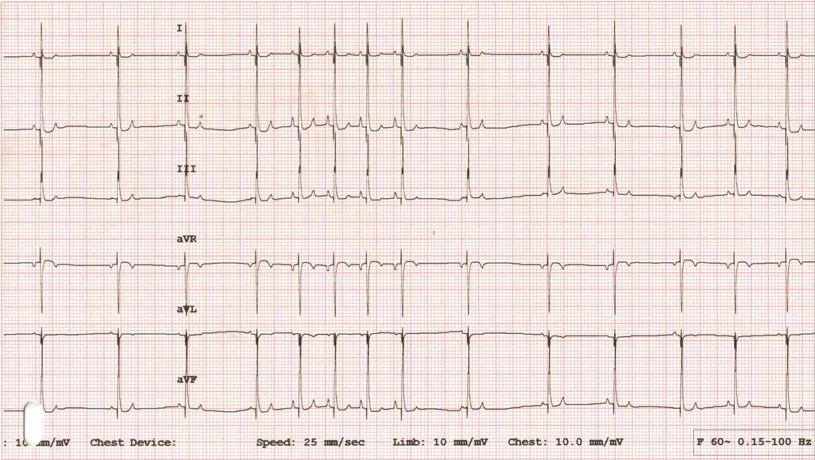
ECG findings
- Rhythm: Regularly irregular, with a cyclical variation in both the R to R intervals and the P to P intervals. There is gradual acceleration of the sinus rate during inspiration, and sudden deceleration during expiration.
- Rate: An average heart rate of around 100 beats per minute, ranging from about 75 beats per minute up to about 120 beats per minute.
- Beansie's ECG.
- PR interval: Within normal limits.
- QRS: Less than 70 ms, so narrow, and consistent with supraventricular activation. QRS morphology is consistent throughout the tracing.
Two things are moving here, and they are moving together. The rate rises and falls in a cycle, and the P waves change shape across that same cycle. Neither of those is a beat arriving early, and neither of them is a beat arriving from the wrong place. That distinction is the whole case.
Respiratory sinus arrhythmia with a wandering pacemaker. In a dog, this represents normal canine physiology, not cardiac conduction disease.
Calculating the rate, when the rate keeps moving.
Here is the practical problem. If you measure a single R to R interval and convert it, you get an instantaneous heart rate, and in this dog that number will be quite different depending on which two beats you happened to pick. So you need an average.
The way I recommend doing that is the pen method.
The pen method
- A ballpoint pen is around 15 centimetres, so 150 millimetres.
- This paper speed is 25 millimetres per second, and 150 divided by 25 is six. So one pen length is six seconds of ECG.
- Lay the pen on the trace and count the QRS complexes along it.
- Multiply that count by 10, because six seconds times 10 is 60 seconds. That gives you beats per minute.
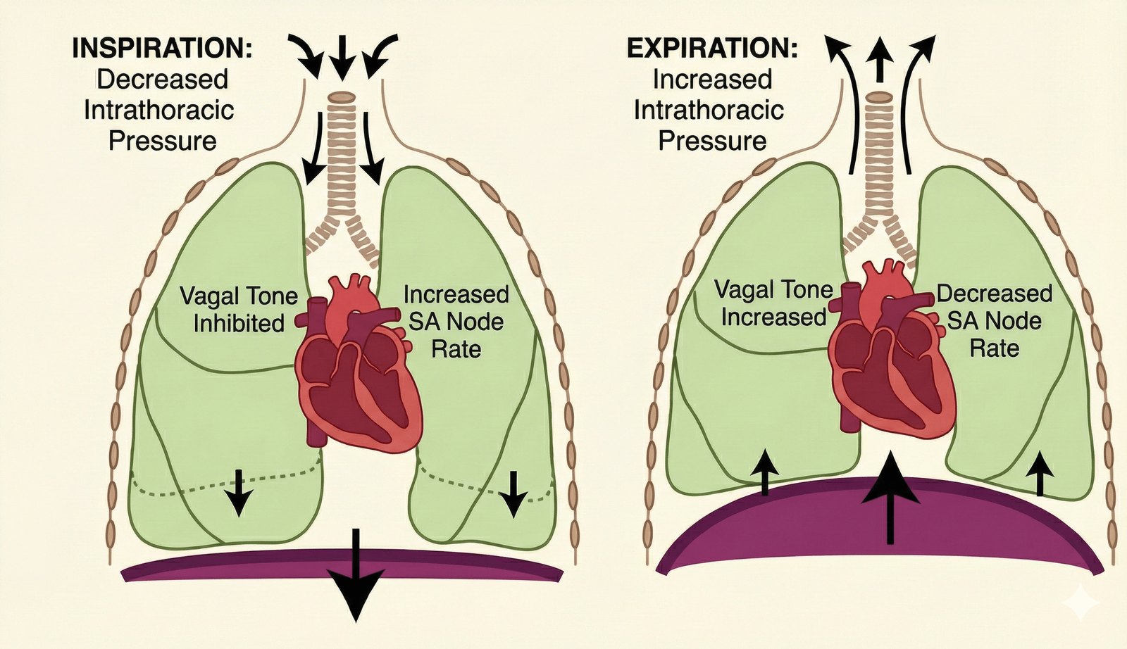
Why the rate follows the breath.
The sinus node is the heart's dominant pacemaker because it has the highest intrinsic automaticity, but its firing rate is continuously modulated by the autonomic nervous system. There are two competing influences. The parasympathetic nervous system slows the heart down, and the sympathetic nervous system speeds it up.
During inspiration the thoracic cavity expands, and that expansion lowers intrathoracic pressure. The pressure drop improves venous return to the heart, so more blood flows into the right atrium. The right atrium stretches slightly, stretch receptors signal to the central nervous system that venous return has increased, and the autonomic nervous system responds by increasing sympathetic tone. That sympathetic input acts directly on the sinus node to increase the heart rate.
On expiration the opposite happens. The thoracic cavity becomes smaller, intrathoracic pressure increases, venous return decreases, the right atrium stretches less. Sympathetic tone withdraws, and that allows the parasympathetic, or vagal, influence to dominate. Parasympathetic tone slows sinus node automaticity, and the heart rate falls.
Why the P waves change shape.
This is the part that gets misread as ectopy, so it is worth being precise about.
The anatomy of the dog's sinus node is a large, elongated, banana-like structure sitting within the high right atrium. Because the sinus node complex is so large, different regions within it can assume pacemaker dominance, and the location of the leading pacemaker site is largely determined by autonomic tone.
Under higher sympathetic tone, such as during inspiration, the leading pacemaker site shifts cranially and superiorly, towards the cranial vena cava. Under high parasympathetic or vagal tone, such as during expiration or at rest, the pacemaker site shifts caudally, more inferiorly, towards the lower atrial floor.
Because atrial depolarization is starting from slightly different locations, the P wave morphology changes from beat to beat. This is a normal shifting of the leading pacemaker site within the sinus node in response to autonomic tone. It is not atrial ectopy.
Differential diagnoses.
These are the rhythms that need ruling out, and the features that rule them out.
| Atrial premature complexes (APCs) | Sick sinus syndrome | Sinoatrial block |
|---|---|---|
| A sudden premature, ectopic beat. An early P′ wave, of different morphology to the sinus P wave, often followed by a non-compensatory pause. | Abrupt sinus pauses, which are not respiration-linked. These patients often have clinical signs. | Pauses usually occur in exact multiples of the P to P interval, rather than the gradual variation in P to P and R to R that we see here. |
Differential diagnoses for respiratory sinus arrhythmia
Clinical significance.
- This is a very common and normal finding, particularly in dogs. It is a marker of normal autonomic tone.
- We typically see it in calm, fit dogs at rest.
- It is not really seen in cats in the hospital, because they have a higher sympathetic tone there, but it can be seen in the home environment and has been documented on Holter recordings.
- If this patient went on to have an echocardiogram, we would expect it to be normal.
And there is a point here that is worth carrying into every other case you see. The presence of respiratory sinus arrhythmia argues against heart failure, where we normally have increased sympathetic tone and often a patient that is tachycardic.
Echo findings.
Management and prognosis.
- No treatment is indicated.
- There are no contraindications to general anesthesia based on these findings.
- Excellent prognosis.
Beansie's echo. A structurally normal heart, which is exactly what a normal variant should look like underneath.
Summary.
- Respiratory sinus arrhythmia and wandering pacemaker are normal canine rhythms.
- They are driven by cyclic changes in autonomic tone during respiration.
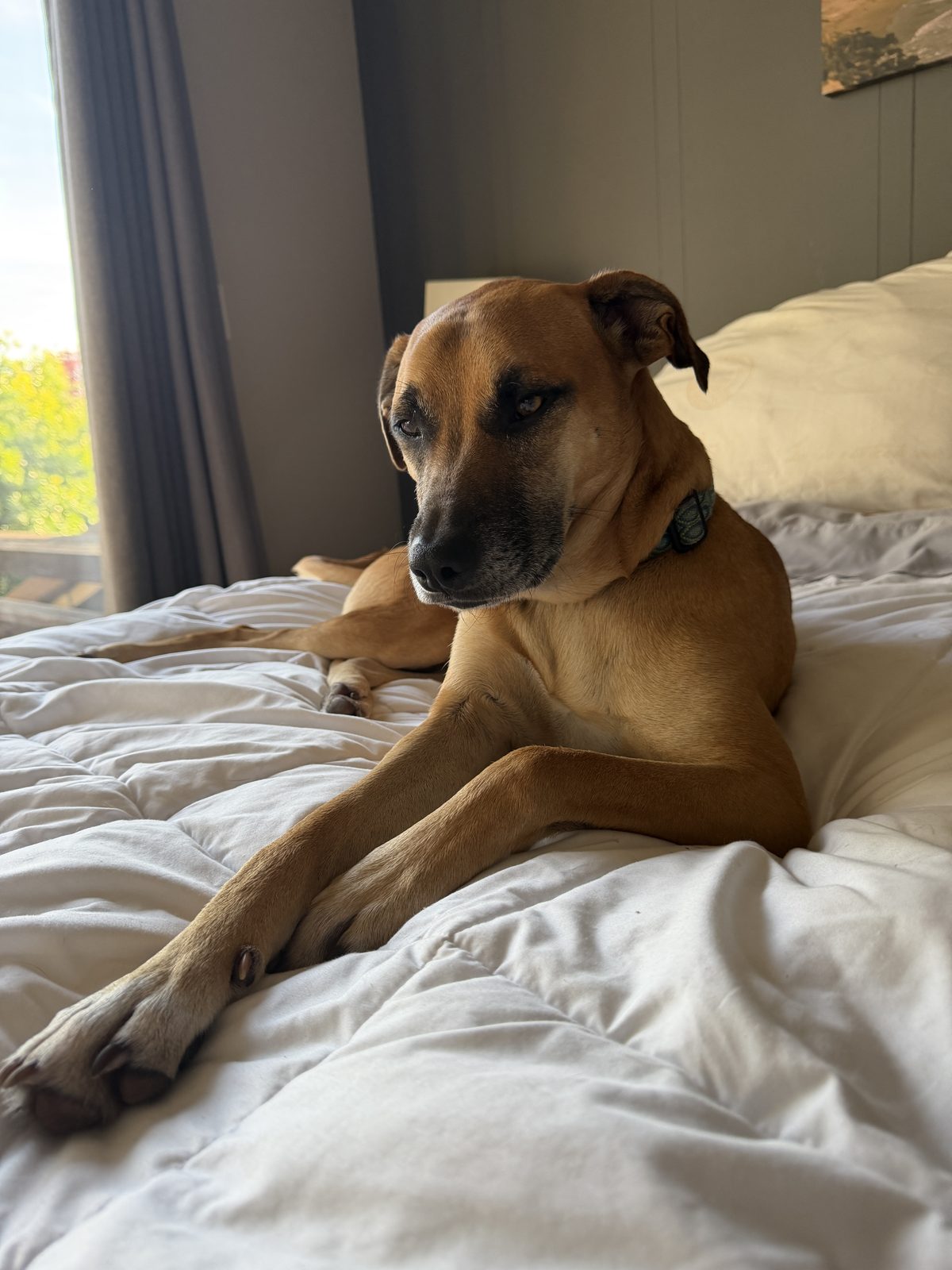
- Beansie left the hospital very happy to have a treat. The owner was happy. Surgery was scheduled, and it all went ahead smoothly.
- Beansie at home. Nice and relaxed, which is more or less the point.
- No treatment is necessary, and there is no contraindication to anesthesia.
Common questions about sinus arrhythmia.
What is respiratory sinus arrhythmia in a dog?
It is a cyclical variation in the sinus rate that follows the respiratory cycle. The rate gradually accelerates during inspiration and decelerates during expiration, so the rhythm sounds and looks regularly irregular. In dogs this is normal physiology and a marker of normal autonomic tone, not cardiac conduction disease.
Is it safe to anesthetize a dog with sinus arrhythmia?
Based on this finding alone, yes. Respiratory sinus arrhythmia with a wandering pacemaker is a normal variant, so no treatment is indicated and there is no contraindication to general anesthesia. The reason to run the ECG is not that the rhythm is dangerous, it is that you need to know which rhythm you are looking at before you anesthetize.
What is a wandering pacemaker?
The canine sinus node is a large, elongated structure in the high right atrium, not a single point. Different regions within it can take over as the leading pacemaker depending on autonomic tone. Under sympathetic tone during inspiration the leading site shifts cranially, giving taller, more peaked P waves. Under vagal tone during expiration it shifts caudally, giving lower voltage P waves. Because depolarization starts from a slightly different place, the P wave changes shape.
How do you tell a wandering pacemaker from atrial ectopy?
Timing. A wandering pacemaker changes the shape of the P wave while the sinus timing is preserved, so the rhythm still varies gradually with the breath. An atrial premature complex is a sudden, early beat, with a P′ wave of different morphology arriving before it was due, and it is often followed by a non-compensatory pause. Variable P wave morphology with preserved sinus timing supports a wandering pacemaker, not ectopy.
How do you calculate heart rate when the rhythm keeps changing?
Take an average rather than an instantaneous rate, because a single R to R interval will give you a very different number depending on where in the respiratory cycle you measured it. The pen method is quick. A ballpoint pen is around 150 mm, and at a paper speed of 25 mm per second that is six seconds of trace. Count the QRS complexes along the pen and multiply by 10.
Do cats get respiratory sinus arrhythmia?
Not usually in the hospital, because their sympathetic tone is higher there. It can be seen in the home environment, and it has been documented on Holter recordings.
Why does sinus arrhythmia argue against heart failure?
Because heart failure is a high sympathetic tone state, and those patients are often tachycardic. Respiratory sinus arrhythmia depends on vagal tone being able to dominate at intervals. If you can see a clear respiratory sinus arrhythmia, that is a strong argument against a heart that is failing.
Get the full case breakdowns
Every case, in full, with the video walkthrough.
Each case breakdown on this site has a longer version. Dr. Hayley works through the ECG and the echo on video, the way she would talk a colleague through it. Subscribers get the full walkthrough with every case.
- The full video walkthrough with each case.
- New case breakdowns as they publish.
- Practical cardiology for vets and techs.
We respect your privacy.Privacy policy·Unsubscribe anytime.